Physical Description
SIZE
Mature Marianina rosea observed on Heron Island reached a maximum of 5mm in length. Adults reach an average length of 6-9mm, and It has been reported that specimens can grow up to 14mm (Marshall & Willan, 1999).
COLOURATION
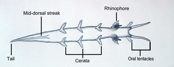
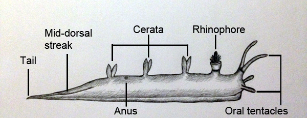
All individuals of M. rosea exhibit a bright magenta colouration across their body, the defining characteristic of which the rosy nudibranch derives its name. White bifurcated and symmetrically paired cerata (dorsal processes) line the dorsal surface of the animal and lead to a white mid dorsal streak that extends through to the tip of the tail (Pruvot-Fol1930; Marshall & Willan, 1999). The basal section of the oral tentacles is magenta, with the remainder of the tentacle exhibiting an opaque white colouration. The basal quarter of the rhinophoral sheath is magenta and the tip ranges from cream to opaque white. The distinctive rhinophoral clavus, consisting of a ring of thin papillae that circle a longer central stalk, is bathed in a rich blood-red colouration (Willan 1988; Marshall & Willan, 1999).
EXTERNAL MORPHOLOGY
Marianina rosea is a small, slender and elongated nudibranch with a high and rounded dorsum. The muscular foot reflects the same width as the mantle, and extends to a long pointed tail. The anterior foot corners are prominent and resemble sharply pointed tentacles (Marshall & Willan, 1999; Cobb & Willan 2006; Debelius & Kuiter 2007). There are two pairs of oral tentacles located on either side of the mouth. The first pair are long, slender and taper to a pointed tip, while the second pair are much shorter in length and display a rounded tip. The chemosensory rhinophores are characteristic of M. rosea, bearing long papillae that resemble an upraised hand (palmate). A tubular sheath displaying simple margins encloses each rhinophore structure. Marianina rosea bear up to four pairs of cerata along the dorsal surface, that aid in gas exchange and absorption of oxygen from the water column. Cerata are bifurcated, smooth and fusiform in structure, with each ceras branching from the main axis and tapering to a pointed tip (Marshall and Willan, 1999). Upon first inspection of M. rosea, individuals are often mistaken for an Aeolid because of their small body size, the presence of long oral tentacles, the presence of antero-lateral tentacular foot corners, and the lack of an oral veil and frontal papillae. The palmate rhinophores, with distinctive sheaths, are the underlying morphological feature of the Tritoniids that places M. rosea into this family.
The anus is located on the right side of the body towards the posterior end of the animal. Approximately one-third of the way down the animal’s dorsal surface is the location of the reproductive aperture. This opening is found on the right side of the body between the rhinophores and first pair of cerata (Behrens 2005).
Dorsal view of Marianina rosea (biological drawing by Elisha Simpson, 2013).

Lateral view of Marianina rosea (biological drawing by Elisha Simpson, 2013).

Juveniles display the bright magenta colouration, however, external morphological structures are generally undeveloped and considerably shorter in length. Juveniles will not reach sexual maturity until they have completed development (Willan 1988).
Slight variations in colour, size and morphology exist between tropical and temperate populations, however, all specimens exhibit the distinctive blood red rhinophoral clavus (Willan & Coleman, 1984). The specimen of M. rosea located at Heron Island was a well-developed specimen of 5mm with only three pairs of cerata present along the dorsal surface of the body. As this specimen displayed well-developed external structures it was unclear as to whether this individual was actually a fully-grown and sexually mature adult due to its small size and only three pairs of cerata present.
|